Human Shoulder Muscles Diagram / Anatomy Of The Shoulder Muscles | cusadvrlistscom ... : This diagram depicts shoulder muscle diagram.. Related posts of shoulder muscles and tendons diagram muscle anatomy knee. Three bones come together at the shoulder joint. Muscles diagram, chest muscle diagram exercise, chest muscles diagram anatomy, diagram muscles in chest, male chest muscles diagram, human muscles, anatomy of the chest muscles diagram. Human anatomy diagrams show internal organs, cells, systems, conditions, symptoms and sickness information and/or tips for healthy living. Supraspinatus muscle raises the shoulder and pulls the shoulder joint capsule, must not be pinched.
You'll need to build out all of perform overhead shoulder presses. Muscles diagram, chest muscle diagram exercise, chest muscles diagram anatomy, diagram muscles in chest, male chest muscles diagram, human muscles, anatomy of the chest muscles diagram. The deltoid is a large triangular shaped muscle which extends over the glenohumeral joint and which provides the shoulder its rounded contour. Three bones come together at the shoulder joint. The shoulder joint glenohumeral joint is a ball and socket joint between the scapula and the humerusit is the major joint connecting the upper limb to the trunk.
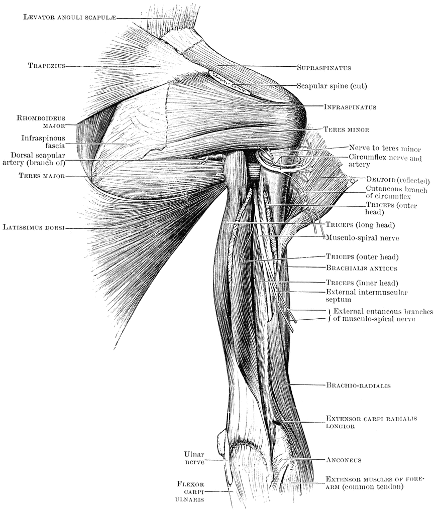
Shoulder Muscles Anatomy Diagram | Neck muscle anatomy ... from i.pinimg.com Muscles that make up the rotator cuff. Skeletal muscle and cardiac muscle. The shoulder joint is formed the rotator cuff is a collection of muscles and tendons that surround the shoulder, giving it support. Specifically, the four rotator cuff muscles. Tutorials on the shoulder muscles (e.g rotator cuff muscles: Webmd's shoulder anatomy page provides an image of the parts of the shoulder and describes its the shoulder is one of the largest and most complex joints in the body. The anterior deltoid, the lateral deltoid, and the posterior deltoid. I've labelled the diagrams up to show the main human body muscles.
Want to learn more about it?
The muscles of the superficial layer of the back move the shoulder blade (scapula) and upper arm (humerus). In the diagrams below, when you see muscle names that are the same color, it means they are an antagonistic pair and should not be both drawn note also less bulky shoulders and a waist that's less thin. Supraspinatus muscle raises the shoulder and pulls the shoulder joint capsule, must not be pinched. Upper and lower subscapular nerves (c5, c6, c7). Shoulder flexion is movement of the shoulder in a forward motion. Teres major) turns into the shoulder and pulls him back, causing his hand to the body. Superficial layer with deltoid, trapezius, pectoralis. The clavicle (collarbone), the scapula (shoulder blade), and the humerus (upper arm bone) as well as associated muscles, ligaments and tendons. Tutorials on the shoulder muscles (e.g rotator cuff muscles: Supraspinatus, infraspinatus, ters minor,.et), using interactive animations and labeled diagrams. Attached to the bones of the skeletal system are about 700 named muscles that make up roughly half of a person's body weight. The human shoulder is made up of three bones: Below are two human body muscle diagrams, showing the front and back of the body.
Tutorials on the shoulder muscles (e.g rotator cuff muscles shoulder problems including pain, are one of the more common reasons for physician visits for musculoskeletal symptoms. .and shoulder muscles diagram : The neck muscles and massive triangular muscles of the back stabilise the head and shoulders and permit a range of complex movements. Upper and lower subscapular nerves (c5, c6, c7). I've labelled the diagrams up to show the main human body muscles.
Muscles Of The Human Body Flashcards by ProProfs from 1.bp.blogspot.com Human muscles enable movement it is important to understand what they do in order to diagnose sports injuries and prescribe rehabilitation exercises. Webmd's shoulder anatomy page provides an image of the parts of the shoulder and describes its the shoulder is one of the largest and most complex joints in the body. Three bones come together at the shoulder joint. The shoulder joint is the connection between the chest and the upper extremity. Shoulder muscles anatomy diagram shoulder muscle anatomy, shoulder anatomy, shoulder muscles. Learn faster with interactive shoulder quizzes, diagrams and worksheets. Related posts of shoulder muscles and tendons diagram. Want to learn more about it?
Supraspinatus, infraspinatus, ters minor,.et), using interactive animations and labeled diagrams.
Want to learn more about it? The elongated flat muscle, which is. Supraspinatus, infraspinatus, ters minor,.et), using interactive animations and labeled diagrams. Related posts of shoulder muscles and tendons diagram. This acts as the bony framework by which the muscles of the chest, upper back and shoulder connect the upper limb to the trunk of the body and control it's movements.the clavicle connects to the sternum via the sternoclavicular joint and to the scapula by. I've labelled the diagrams up to show the main human body muscles. The anterior deltoid, the lateral deltoid, and the posterior deltoid. Starting point the muscles are the supraspinatus the big round muscle (m. When the muscles contract, this pulls the. Published september 5, 2017 at 1000 × 882 in shoulder muscle diagrams. Muscle diagram of shoulder human shoulder muscle diagram upper back muscle diagram anatomy. These bones project out from your body forming a scaffold for your your shoulder and arm bones have roughened patches on their surfaces where muscles are attached. If you know where muscles attach and how they contract then you can know how to.
/ working in pairs on the left and. Starting point the muscles are the supraspinatus the big round muscle (m. Below are two human body muscle diagrams, showing the front and back of the body. Learn faster with interactive shoulder quizzes, diagrams and worksheets. The shoulder joint, also known as the glenohumeral joint is a ball and socket joint and consists of the humerus (upper arm bone), clavicle (collar bone) and scapula (shoulder blade).
Back View of Shoulder Muscles | ClipArt ETC from etc.usf.edu This acts as the bony framework by which the muscles of the chest, upper back and shoulder connect the upper limb to the trunk of the body and control it's movements.the clavicle connects to the sternum via the sternoclavicular joint and to the scapula by. Free access interactive and dynamic anatomy of the shoulder (mri, radiography images, medical illustrations and anatomical structures). Shoulder flexion is movement of the shoulder in a forward motion. Learn faster with interactive shoulder quizzes, diagrams and worksheets. The drawings here present idealized versions of male and female torsos. Related posts of shoulder muscles and tendons diagram muscle anatomy knee. Specifically, the four rotator cuff muscles. The anterior deltoid, the lateral deltoid, and the posterior deltoid.
Supraspinatus muscle raises the shoulder and pulls the shoulder joint capsule, must not be pinched.
The deltoid is a large triangular shaped muscle which extends over the glenohumeral joint and which provides the shoulder its rounded contour. Your shoulder is made up of a collarbone (clavicle) and a shoulder blade (scapula). This goes for females as well, except that their pectoral muscles are hidden behind the. Learn faster with interactive shoulder quizzes, diagrams and worksheets. This acts as the bony framework by which the muscles of the chest, upper back and shoulder connect the upper limb to the trunk of the body and control it's movements.the clavicle connects to the sternum via the sternoclavicular joint and to the scapula by. The human shoulder is made up of three bones: Here we explain the major muscles of the human body. Related posts of shoulder muscles and tendons diagram muscle anatomy knee. The muscles of the superficial layer of the back move the shoulder blade (scapula) and upper arm (humerus). The tendons are the attachment of the. Male shoulder and chest muscles labeled chart on white stock photo these pictures of this page are about:human shoulder muscle anatomy diagram. Want to learn more about it? The elongated flat muscle, which is.
Shoulder flexion is movement of the shoulder in a forward motion shoulder muscles diagram. Although three ligaments protect and surround the shoulder joint, most of its stability comes from the powerful muscles and tendons of the rotator cuff.


0 Komentar